Tendon Diagram : The main portion inserts into the tuberosity of the navicular bone.. Muscles that are not used can get smaller and weaker true. Sprains happen when ligaments (which connect bones to bones) are stretched too much. The tendon of the tibialis posterior muscle (sometimes called the posterior tibial tendon) descends posterior to the medial malleolus. Rotator cuff injuries are very common, affecting over 3 million people in the united states every year. Gliding now medially shows the insertion of the subscapularis tendon.
The main portion inserts into the tuberosity of the navicular bone. The tendon of the tibialis posterior muscle (sometimes called the posterior tibial tendon) descends posterior to the medial malleolus. If a muscle is not used, it will get smaller. Let us have a detailed overview of the animal cell, its types, diagram and structure. A sprain happens when a tendon is stretched too much.
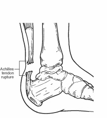
Achilles Tendon Rupture - Foot Health Facts from www.foothealthfacts.org If you would like to learn all the parts of the foot structure, you have come to the right place. Jul 30, 2018 · diaphragm diagram. The femur is the only bone of the thigh. If a muscle is not used, it will get smaller. Let us have a detailed overview of the animal cell, its types, diagram and structure. Rotator cuff injuries are very common, affecting over 3 million people in the united states every year. A sprain happens when a tendon is stretched too much. The main portion inserts into the tuberosity of the navicular bone.
Posterior tibial tendon dysfunction (pttd) is a condition caused by changes in the tendon, impairing its ability to support the arch.
Gliding now medially shows the insertion of the subscapularis tendon. The femur is the only bone of the thigh. If a muscle is not used, it will get smaller. Most people with rotator cuff injuries can recover with rest and physical therapy. It terminates by dividing into plantar, main, and recurrent components. This results in flattening of the foot. The subscapularis tendon lies approximately 3 to 5 cm under the surface. A retinaculum refers to any region on the body in which tendon groups from different muscles. A stretching injury to a tendon (which connects a muscle to a bone) is called a strain. Rotator cuff injuries are very common, affecting over 3 million people in the united states every year. Longitudinal plane of the musculus subscapularis and its tendon. The thigh and leg bones articulate at the knee joint that is protected and enhanced by the patella bone that supports the quadriceps tendon. Sprains happen when ligaments (which connect bones to bones) are stretched too much.
Most people with rotator cuff injuries can recover with rest and physical therapy. Quite deep for ultrasonography, and therefore displaying through a highly penetrative 5 mhz linear applicator is worth a try. A stretching injury to a tendon (which connects a muscle to a bone) is called a strain. Longitudinal plane of the musculus subscapularis and its tendon. Muscles that are not used can get smaller and weaker true.
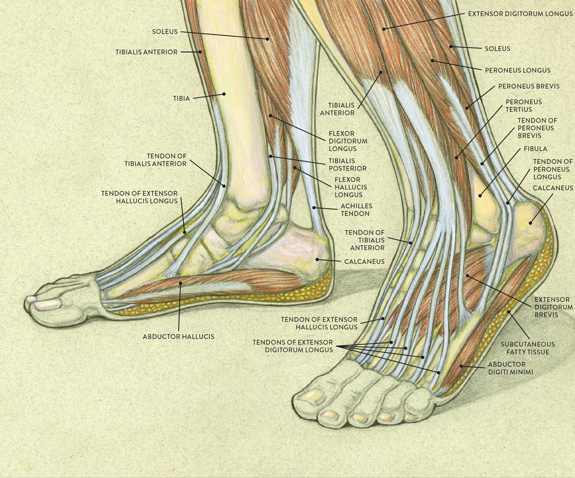
Foot Anatomy Tendons : Muscles Of The Foot Dorsal Plantar ... from doctorlib.info A retinaculum refers to any region on the body in which tendon groups from different muscles. If a muscle is not used, it will get smaller. Quite deep for ultrasonography, and therefore displaying through a highly penetrative 5 mhz linear applicator is worth a try. Sprains happen when ligaments (which connect bones to bones) are stretched too much. Most people with rotator cuff injuries can recover with rest and physical therapy. If you would like to learn all the parts of the foot structure, you have come to the right place. A stretching injury to a tendon (which connects a muscle to a bone) is called a strain. Let us have a detailed overview of the animal cell, its types, diagram and structure.
The thigh and leg bones articulate at the knee joint that is protected and enhanced by the patella bone that supports the quadriceps tendon.
Sprains happen when ligaments (which connect bones to bones) are stretched too much. Rotator cuff injuries are very common, affecting over 3 million people in the united states every year. If you would like to learn all the parts of the foot structure, you have come to the right place. A sprain happens when a tendon is stretched too much. Quite deep for ultrasonography, and therefore displaying through a highly penetrative 5 mhz linear applicator is worth a try. The femur is the only bone of the thigh. The subscapularis tendon lies approximately 3 to 5 cm under the surface. Let us have a detailed overview of the animal cell, its types, diagram and structure. Longitudinal plane of the musculus subscapularis and its tendon. Posterior tibial tendon dysfunction (pttd) is a condition caused by changes in the tendon, impairing its ability to support the arch. A retinaculum refers to any region on the body in which tendon groups from different muscles. Jul 05, 2018 · the foot diagram has a complex structure made up of bones, ligaments, muscles, and tendons. However, more serious injuries, such as complete rotator cuff tears, may require surgical repair.
Most people with rotator cuff injuries can recover with rest and physical therapy. Rotator cuff injuries are very common, affecting over 3 million people in the united states every year. Let us have a detailed overview of the animal cell, its types, diagram and structure. Jul 05, 2018 · the foot diagram has a complex structure made up of bones, ligaments, muscles, and tendons. A stretching injury to a tendon (which connects a muscle to a bone) is called a strain.
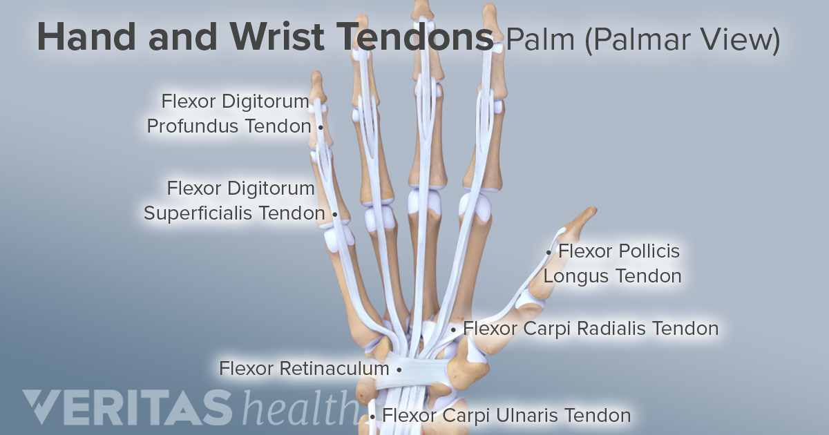
Ligaments, Tendons, and Nerves of the Wrist from embed.widencdn.net Most people with rotator cuff injuries can recover with rest and physical therapy. Muscles that are not used can get smaller and weaker true. The tendon of the tibialis posterior muscle (sometimes called the posterior tibial tendon) descends posterior to the medial malleolus. Rotator cuff injuries are very common, affecting over 3 million people in the united states every year. Jul 30, 2018 · diaphragm diagram. The femur is the only bone of the thigh. This results in flattening of the foot. Professor geoffrey meyer bsc (hons) phd frsb.
This results in flattening of the foot.
The subscapularis tendon lies approximately 3 to 5 cm under the surface. A stretching injury to a tendon (which connects a muscle to a bone) is called a strain. Posterior tibial tendon dysfunction (pttd) is a condition caused by changes in the tendon, impairing its ability to support the arch. Understanding the structure of the foot is best done by looking at a foot diagram where the anatomy has been labeled. The tendon of the tibialis posterior muscle (sometimes called the posterior tibial tendon) descends posterior to the medial malleolus. Muscles that are not used can get smaller and weaker true. Jul 30, 2018 · diaphragm diagram. This results in flattening of the foot. A sprain happens when a tendon is stretched too much. However, more serious injuries, such as complete rotator cuff tears, may require surgical repair. Rotator cuff injuries are very common, affecting over 3 million people in the united states every year. Jul 05, 2018 · the foot diagram has a complex structure made up of bones, ligaments, muscles, and tendons. Quite deep for ultrasonography, and therefore displaying through a highly penetrative 5 mhz linear applicator is worth a try.


0 Komentar